Hemochromatosis and Bone Loss: When Excess Iron Damages Bone
At the opposite end of the iron spectrum from deficiency, iron overload — most commonly from hereditary hemochromatosis — produces an under-recognized form of secondary osteoporosis. The condition affects roughly 1 in 200-300 people of northern European descent (one of the most common genetic disorders) and is often diagnosed late. By the time it is recognized, bone density loss has often progressed.
What hemochromatosis does to bone
Excess iron accumulates in osteoblasts and the bone matrix, where it inhibits collagen synthesis and crosslinking. It also contributes to hypogonadism — iron deposition in the pituitary reduces gonadotropin release, which lowers testosterone and estrogen. Lower sex hormones drive accelerated bone loss. Both pathways converge to produce osteoporosis prevalence of 25-40% in untreated hemochromatosis patients.
How it presents
Hemochromatosis often presents nonspecifically: fatigue, joint pain, abnormal liver enzymes, sometimes bronzed skin. The osteoporosis is rarely the presenting feature, but it tracks with other manifestations of the disease. Joint pain that turns out to be hemochromatosis-related arthropathy is a common finding that prompts diagnosis.
Who should be screened
Family history of hemochromatosis. Personal history of unexplained elevated transaminases, joint pain, fatigue, or diabetes (especially with relatively normal weight). Postmenopausal women or men of any age with unexplained osteoporosis (not a typical age or risk profile). Screening: serum iron studies (ferritin over 300 ng/mL in men or 200 ng/mL in women, with elevated transferrin saturation over 45%) prompts genetic testing for HFE gene mutations.
Treatment and bone effects
Treatment is therapeutic phlebotomy — regular blood removal to deplete iron stores. With sustained treatment, ferritin normalizes within 6-18 months. Bone density partially recovers, though the recovery is incomplete in older patients. Standard osteoporosis treatments (bisphosphonates, denosumab) are also commonly used to address the established bone loss.
Other iron-overload causes
Beyond hereditary hemochromatosis, iron overload occurs in thalassemia, sickle cell disease, transfusion-dependent anemias, and some forms of myelodysplastic syndrome. Each requires iron chelation therapy and parallel bone-health management. The bone-density effect is similar across causes.
Monitoring after diagnosis
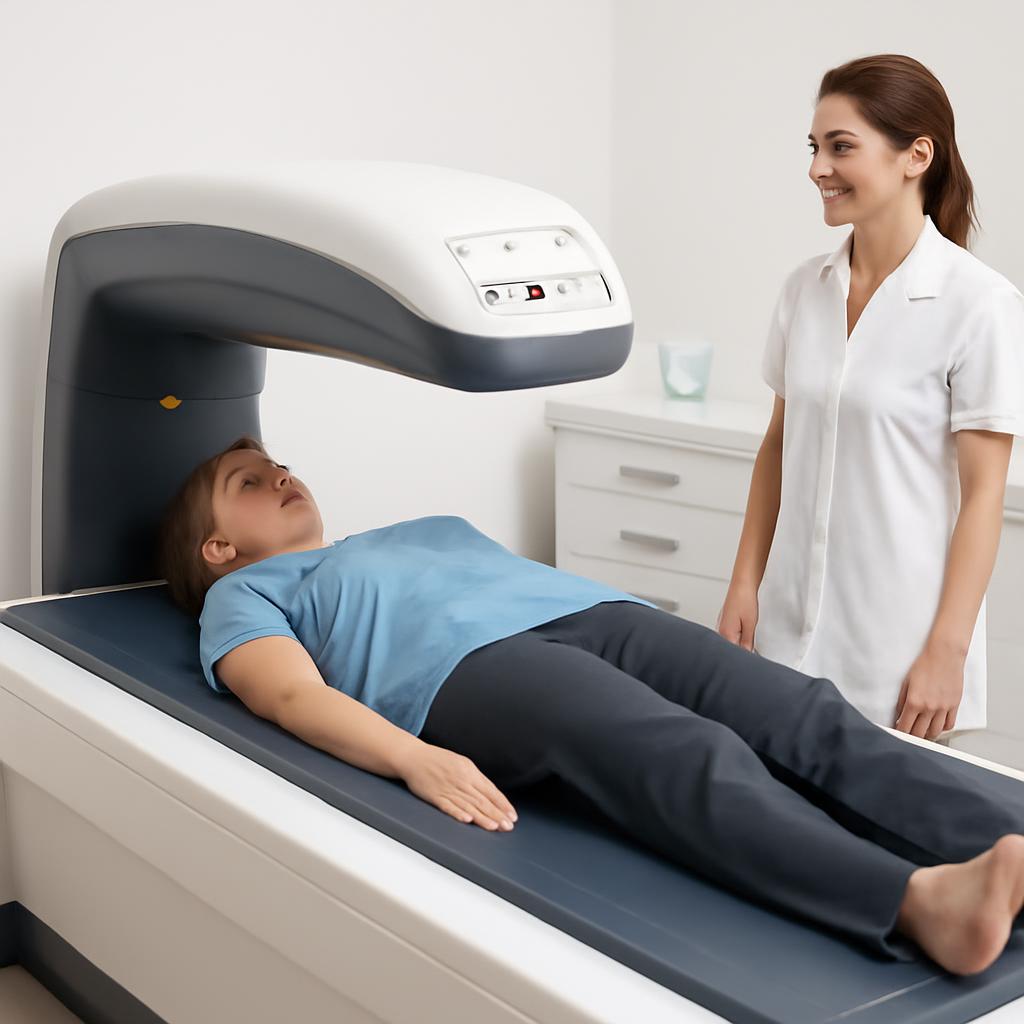
DEXA at diagnosis and then every 1-2 years until stable. Annual hormone testing (testosterone in men, estrogen and FSH in women if perimenopausal). Continued bone-supportive measures: calcium, vitamin D, K2, resistance training, weight-bearing exercise. The interventions are familiar; the underlying condition adds urgency.
Frequently asked questions
How common is hemochromatosis?
The HFE gene mutation is one of the most common genetic mutations in northern European populations. Clinically expressed disease is less common but still affects roughly 1 in 200-300 people of that ancestry.
Is the bone loss reversible with phlebotomy?
Partially. Phlebotomy normalizes the active iron-overload contribution but does not fully recover the bone density already lost. Standard osteoporosis treatment is often added.
Should I worry about iron supplementation causing hemochromatosis?
Iron supplementation does not cause hemochromatosis — the genetic mutation does. But people with hemochromatosis must avoid iron supplements and limit dietary iron, particularly heme iron from red meat.
Can blood donation help?
Yes — regular blood donation is essentially the same as therapeutic phlebotomy. Many blood centers now accept donations from hemochromatosis patients specifically for this reason.
Related reading: The Bone Density Solution review · Vitamin K2 and bone health